Advanced Technology Used
at Lauren Alexander Vision Source
OPTOS Retinal Exam
Annual eye exams are vital to maintaining your vision and overall health. We use cutting-edge digital imaging technology to evaluate the health of your eyes. This is crucial as many eye diseases can be treated successfully if detected early on.
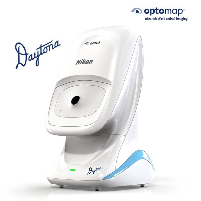
To that end, we offer the optomap® Retinal Exam as an important part of your comprehensive eye exam. The optomap® Retinal Exam produces an image that is as unique as your fingerprint and provides a wide image to examine the health of your retina.
Many eye problems can develop without you knowing, and you may not notice any change in your sight. However, diseases such as macular degeneration, glaucoma, retinal tears or detachments, and other health problems such as diabetes and high blood pressure can be detected with a retinal eye exam. The earlier the detection and treatment, the more effective the outcome.
The optomap® Retinal Exam is fast, easy, and comfortable for people of all ages. Simply look into the device one eye at a time and experience a comfortable flash of light that will let you know the image of your retina has been taken. The optomap® image is immediately displayed on a computer screen for your eye doctor to review it with you.

Corneal Mapping
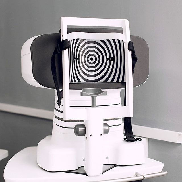
Corneal topography, also known as photokeratoscopy or videokeratography, is a diagnostic tool that provides 3-D images of the cornea. A corneal topography test delivers detailed 3D maps of the cornea’s shape and curvature and helps detect corneal diseases and irregular corneal conditions, such as swelling, scarring, abrasions, deformities, and irregular astigmatisms.
Corneal topography can be used for the following:
Diagnosing and monitoring Keratoconus
Planning refractive surgery
Monitoring eye health following refractive surgery
Determining appropriate intraocular lens for cataract surgery
Evaluating and treating astigmatism following keratoplasty
Detecting corneal conditions such as pterygia, corneal scars, and Salzmann nodules
Monitoring eye diseases
Examining corneal endothelium
Glaucoma: assessing the integrity of the anterior angle
Measuring corneal depth
Evaluating corneal nerve
Detecting infectious keratitis

Digital Retinal Imaging
Retinal imaging takes a digital picture of the back of your eye and displays the retina, the optic disk, and blood vessels on the computer screen. This image is electronically stored, giving the eye doctor a permanent record of the state and health of your retina.
Any changes to your retina will be detected each time you get your eyes examined. This is crucial, as many eye conditions, such as glaucoma, diabetic retinopathy, and macular degeneration are diagnosed by detecting changes over time.
To conduct the eye exam, your eye doctor may dilate your eyes using special drops to widen your pupils. Next, you'll place your chin and forehead on a support, open your eyes as wide as possible, and stare straight ahead while a laser scans your eyes.
If your eyes have been dilated, you'll experience blurred vision and light sensitivity for about 4 hours. Wear sunglasses and have someone drive you home after your eye exam.
Optical Coherence Tomography (OCT)
An Optical Coherence Tomography scan (or OCT scan) is a non-invasive imaging test. This technology uses light waves to take cross-section pictures of your retina.
With OCT, your eye doctor can see color-coded, cross-sectional images of the retina, and can map and measure each layer's thickness. These measurements help diagnose and treat retinal diseases, such as age-related macular degeneration (AMD), glaucoma, retinal detachment, and diabetic retinopathy.
An OCT scan is a non-invasive, painless test that takes only 10 minutes to complete. We invite you to contact our office to inquire about getting an OCT at your next appointment

Visual Field Testing
As you read the words on this page, how much can you see out of the corners of your eyes? Can you see what's happening around you?
Your visual field refers to how wide of an area your eye can see when you concentrate on a single point. Visual field testing allows your eye doctor to determine how much vision you have in either eye, and how much vision loss may have occurred over time.
A visual field test helps determine whether you have blind spots (scotoma) in your vision and where they are located. In those with glaucoma, the visual field test helps to establish any possible peripheral vision loss resulting from this disease.
Eye doctors may also use visual field tests to examine limited vision due to problems such as ptosis and droopy eyelids.
Patients with the following conditions should be monitored regularly for visual field loss:
Glaucoma
Multiple sclerosis
Thyroid eye disease (Graves' disease)
Pituitary gland disorders
Central nervous system problems (such as a tumor that may be pressing on visual parts of the brain)
Stroke
The use of certain medications
If your visual field is limited, driving may be a hazard. Concerned about your vision? Talk with one of the Lauren Alexander Vision Source eye doctors. We can help.


